Devenez membre d'Incathlab et bénéficiez d'un accès complet !
Inscription Connexion
This video presents a clinical case of a 89-year-old female patient with significant aortoiliac disease, characterized by a calcific cauliflower-shaped stenosis in the left common iliac artery (LCIA) and left common femoral artery (LCFA) calcific occlusion treated with the Intravascular Lithotripsy System (Shockwave Medical).
Educational Objectives
- Understand the principles and mechanics of IVL, including how acoustic shockwaves disrupt calcific plaque while preserving surrounding vascular tissue.
- Identify the specific clinical scenarios in which intravascular lithotripsy is indicated, particularly in cases of heavily calcified lesions that may not respond well to traditional angioplasty.
- Explain the procedural steps involved in IVL, angioplasty and stenting for calcific stenosis.
Step-by-step procedure:
Using the Seldinger technique, a 7Fr sheath was inserted and crossover was performed.
A diagnostic catheter was inserted to perform a selective angiography of the aortoiliac region.
The angiography images were used to evaluate the extent of the disease and the morphology of the stenoses.
After angiographic evaluation of the lesions, a 0.014” guidewire was used in order to navigate through the stenoses.
After successfully passing the stenoses, the wire was exchanged to a 0.014” superstiff wire.
Appropriate intravascular lithotripsy (IVL) catheters designed for the vessel sizes were selected.
The catheters were connected to the Shockwave Medical lithotripsy generator.
The IVL catheters were carefully positioned to the site of the stenoses, ensuring proper positioning across the lesions.
The lithotripsy generator was activated to deliver a series of pulses of sonic waves. These shockwaves disrupted the calcific plaques while sparing the surrounding tissue.
Finally, an appropriately sized balloon-expandable stent was selected and placed based on the measured diameter of the LCIA and the characteristics of the stenosis.

Dernière mise à jour : 29/10/2024
Our Cases of the Month
The case of the month is a new way for our users to watch, learn, and share with incathlab. They can watch a video that highlights an innovative case and uses excellent pedagogical techniques, lear...
Suggestions
San Francisco : Lundi 18 septembre 2023 de 01h07 à 01h07 (GMT+2)
New York : Lundi 18 septembre 2023 de 04h07 à 04h07 (GMT+2)
Buenos Aires : Lundi 18 septembre 2023 de 05h07 à 05h07 (GMT+2)
Reykjavik : Lundi 18 septembre 2023 de 08h07 à 08h07 (GMT+2)
London / Dublin : Lundi 18 septembre 2023 de 09h07 à 09h07 (GMT+2)
Paris / Berlin : Lundi 18 septembre 2023 de 10h07 à 10h07 (GMT+2)
Istanbul : Lundi 18 septembre 2023 de 11h07 à 11h07 (GMT+2)
Moscou / Dubaï : Lundi 18 septembre 2023 de 12h07 à 12h07 (GMT+2)
Bangkok : Lundi 18 septembre 2023 de 15h07 à 15h07 (GMT+2)
Shanghai : Lundi 18 septembre 2023 de 16h07 à 16h07 (GMT+2)
Tokyo : Lundi 18 septembre 2023 de 17h07 à 17h07 (GMT+2)
Sydney : Lundi 18 septembre 2023 de 19h07 à 19h07 (GMT+2)
Wellington : Lundi 18 septembre 2023 de 21h07 à 21h07 (GMT+2)
Complex multivascular patient with occluded brachiocephalic trunk
Case of the month: September 2023
San Francisco : Mardi 27 avril 2021 de 07h à 08h (GMT+2)
New York : Mardi 27 avril 2021 de 10h à 11h (GMT+2)
Buenos Aires : Mardi 27 avril 2021 de 11h à 12h (GMT+2)
Reykjavik : Mardi 27 avril 2021 de 14h à 15h (GMT+2)
London / Dublin : Mardi 27 avril 2021 de 15h à 16h (GMT+2)
Paris / Berlin : Mardi 27 avril 2021 de 16h à 17h (GMT+2)
Istanbul : Mardi 27 avril 2021 de 17h à 18h (GMT+2)
Moscou / Dubaï : Mardi 27 avril 2021 de 18h à 19h (GMT+2)
Bangkok : Mardi 27 avril 2021 de 21h à 22h (GMT+2)
Shanghai : Mardi 27 avril 2021 de 22h à 23h (GMT+2)
Tokyo : Mardi 27 avril 2021 de 23h à 00h (GMT+2)
Sydney : Mercredi 28 avril 2021 de 01h à 02h (GMT+2)
Wellington : Mercredi 28 avril 2021 de 03h à 04h (GMT+2)
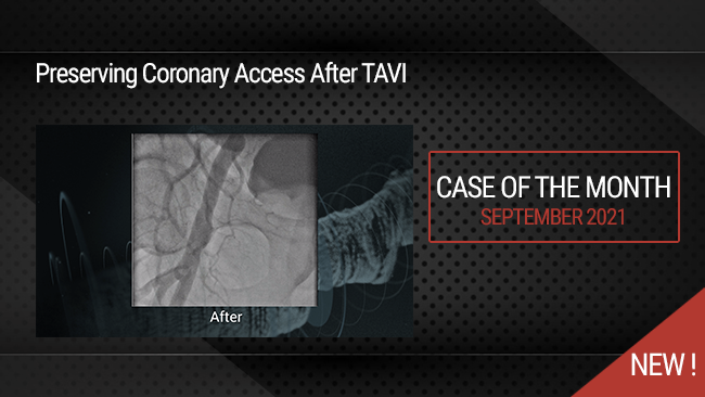
Preserving Coronary Access After TAVI
Case of the month: September 2021
San Francisco : Vendredi 29 juin 2018 de 01h40 à 03h (GMT+2)
New York : Vendredi 29 juin 2018 de 04h40 à 06h (GMT+2)
Buenos Aires : Vendredi 29 juin 2018 de 05h40 à 07h (GMT+2)
Reykjavik : Vendredi 29 juin 2018 de 08h40 à 10h (GMT+2)
London / Dublin : Vendredi 29 juin 2018 de 09h40 à 11h (GMT+2)
Paris / Berlin : Vendredi 29 juin 2018 de 10h40 à 12h (GMT+2)
Istanbul : Vendredi 29 juin 2018 de 11h40 à 13h (GMT+2)
Moscou / Dubaï : Vendredi 29 juin 2018 de 12h40 à 14h (GMT+2)
Bangkok : Vendredi 29 juin 2018 de 15h40 à 17h (GMT+2)
Shanghai : Vendredi 29 juin 2018 de 16h40 à 18h (GMT+2)
Tokyo : Vendredi 29 juin 2018 de 17h40 à 19h (GMT+2)
Sydney : Vendredi 29 juin 2018 de 19h40 à 21h (GMT+2)
Wellington : Vendredi 29 juin 2018 de 21h40 à 23h (GMT+2)

