×
Il semble que vous utilisiez une version obsolète de internet explorer. Internet explorer n'est plus supporté par Microsoft depuis fin 2015. Nous vous invitons à utiliser un navigateur plus récent tel que Firefox, Google Chrome ou Microsoft Edge.
My Player placeholder

Devenez membre d'Incathlab et bénéficiez d'un accès complet !

Vous devez être membre pour accéder aux vidéos Incathlab sans limitation. Inscrivez vous gratuitement en moins d'une minute et accédez à tous les services Incathlab ! Vous avez aussi la possibilité de vous connecter directement avec votre compte facebook ou twitter en cliquant sur login en haut à droite du site.

Inscription Connexion


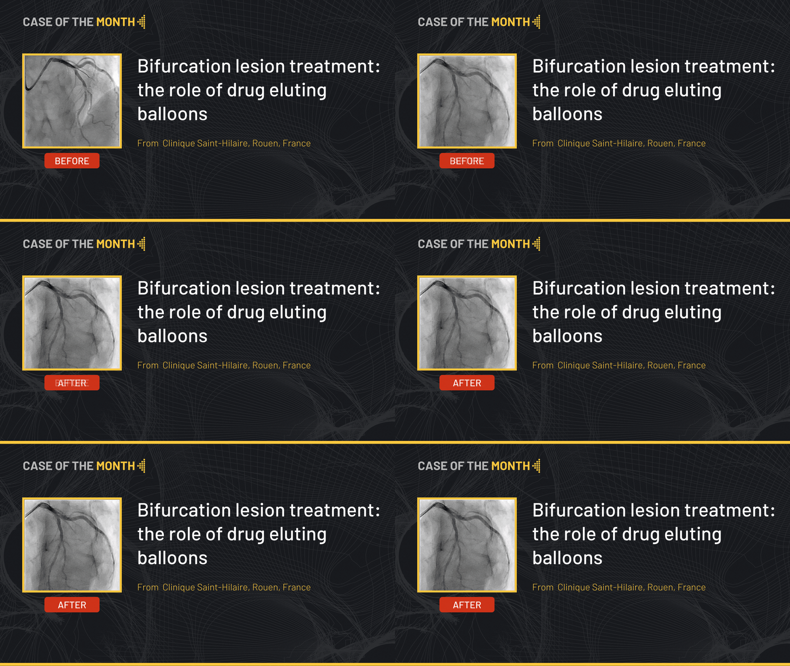
This month’ case highlights the treatment of a bifurcation lesion in a 70 years old female patient, ex-smoker, known to have essential hypertension and chronic obstructive lung disease. The patient reports exertional palpitations for which a coronary computed tomography angiography showed a left anterior descending (LAD) lesion and a Holter ECG multiple ventricular extrasystoles. She also is scheduled for elective surgery.

Coronary angiography pre-TAVR revealed: Severe proximal LAD bifurcation lesion with a relatively small diagonal in an acute angle that covers an important an important territory, Medina (1,1,1).

Educational objectives

  • How to treat a bifurcation lesion
  • Plan a step-by-step approach procedure for bifurcation lesions with a bail-out strategy
  • Role of drug coated balloons in bifurcation lesions
  • Lesion preparation using a scoring balloon
  • Avoiding intimal disruption and side branch occlusion

Step-by-step procedure:

1) Access site:

  • Right radial approach: 6 French EBU 3.5 6 French to the left main.
  • Anticoagulation using heparin.

2) Lesion preparation:

  • Two 0.014” workhorse guidewires were introduced into the LAD and the diagonal respectively.
  • Using the two markers of a non-compliant balloon, calcifications could be seen using enhanced stent imaging.
  • The balloon is inflated at moderate pressure (13 atm) for a longer time in order to undergo a controlled angioplasty and preserve intimal integrity by avoiding dissections.

3) Calcium modification device (scoring balloon):

  • Angiographic assessment of the lesion showed the need for a further dilation using a Calcium modification device.
  • A 2.5 x 13 mm scoring balloon (NSE Alpha) was inflated at 8 atm in order to disrupt the lesion calcium.

4) Treatment decision for the LAD lesion : DCB or DES ?

With the risk of losing the diagonal branch by stenting the LAD, a bail out DK-Crush or V-Stenting techniques could be used. A Drug Coated Balloon would therefore overcome this caging affect as well as the need for longer dual antiplatelet therapy.

A 2.5 x 15 mm Sirolimus drug coated balloon SeQuent SCB was inflated to 10 atm during 45 seconds. Further lesion work or intracoronary imaging should be avoided in order not to disrupt the implanted coating unless a dissection is present.

Final angiographic end-result showed a satisfactory LAD result with the diagonal branch preserved.

Phone call follow-up after 3 months : the patient is fine


Bibliography

1. Corballis, N.H.; Paddock, S.; Gunawardena, T.; Merinopoulos, I.; Vassiliou, V.S.; Eccleshall, S.C. Drug Coated Balloons for Coronary Artery Bifurcation Lesions: A Systematic Review and Focused Meta-Analysis. PLoS One 2021, 16, 1–10, doi:10.1371/journal.pone.0251986.
 
2. Muramatsu, T.; Kozuma, K.; Tanabe, K.; Morino, Y.; Ako, J.; Nakamura, S.; Yamaji, K.; Kohsaka, S.; Amano, T.; Kobayashi, Y.; et al. Clinical Expert Consensus Document on Drug-Coated Balloon for Coronary Artery Disease from the Japanese Association of Cardiovascular Intervention and Therapeutics. Cardiovasc. Interv. Ther. 2023, 38, 166–176, doi:10.1007/s12928-023-00921-2.
 
3. Scheller, B.; Mangner, N.; Abdul Kader, M.A.S.K.; Wan Ahmad, W.A.; Jeger, R.; Wöhrle, J.; Ong, T.K.; Liew, H.B.; Gori, T.; Mahfoud, F.; et al. Combined Analysis of Two Parallel Randomized Trials of Sirolimus-Coated and Paclitaxel-Coated Balloons in Coronary In-Stent Restenosis Lesions. Circ. Cardiovasc. Interv. 2022, 15, E012305, doi:10.1161/CIRCINTERVENTIONS.122.012305.
 
4. de la Torre Hernandez, J.M.; Puri, R.; Alfonso, F. Drug-Coated Balloon: “Scoring to Win.” JACC Cardiovasc. Interv. 2017, 10, 1341–1343, doi:10.1016/j.jcin.2017.05.019.


Date du tournage : 01/03/2024
Dernière mise à jour : 05/03/2024

Our Cases of the Month

The case of the month is a new way for our users to watch, learn, and share with incathlab. They can watch a video that highlights an innovative case and uses excellent pedagogical techniques, lear...

Partager
1 comment
Participer à la discussion
  • Joshua W. Very interesting case.

      Sélectionnez vos fichiers, cliquez sur le bouton envoyer, rédigez votre comentaire puis cliquez sur le bouton envoyer. (formats autorisés : images jpeg, gif, png, and PDF)
      Your browser doesn't have Flash, Silverlight or HTML5 support.


    Suggestions

    Discover your Monthly Case
    Honolulu : Dimanche 17 septembre 2023 de 22h07 à 22h07 (GMT+2)
    San Francisco : Lundi 18 septembre 2023 de 01h07 à 01h07 (GMT+2)
    New York : Lundi 18 septembre 2023 de 04h07 à 04h07 (GMT+2)
    Buenos Aires : Lundi 18 septembre 2023 de 05h07 à 05h07 (GMT+2)
    Reykjavik : Lundi 18 septembre 2023 de 08h07 à 08h07 (GMT+2)
    London / Dublin : Lundi 18 septembre 2023 de 09h07 à 09h07 (GMT+2)
    Paris / Berlin : Lundi 18 septembre 2023 de 10h07 à 10h07 (GMT+2)
    Istanbul : Lundi 18 septembre 2023 de 11h07 à 11h07 (GMT+2)
    Moscou / Dubaï : Lundi 18 septembre 2023 de 12h07 à 12h07 (GMT+2)
    Bangkok : Lundi 18 septembre 2023 de 15h07 à 15h07 (GMT+2)
    Shanghai : Lundi 18 septembre 2023 de 16h07 à 16h07 (GMT+2)
    Tokyo : Lundi 18 septembre 2023 de 17h07 à 17h07 (GMT+2)
    Sydney : Lundi 18 septembre 2023 de 19h07 à 19h07 (GMT+2)
    Wellington : Lundi 18 septembre 2023 de 21h07 à 21h07 (GMT+2)

    Complex multivascular patient with occluded brachiocephalic trunk

    Case of the month: September 2023

    Partager
    Vendredi 29 juin 2018 de 09h40 à 11h (GMT+2)
    Honolulu : Jeudi 28 juin 2018 de 22h40 à 00h (GMT+2)
    San Francisco : Vendredi 29 juin 2018 de 01h40 à 03h (GMT+2)
    New York : Vendredi 29 juin 2018 de 04h40 à 06h (GMT+2)
    Buenos Aires : Vendredi 29 juin 2018 de 05h40 à 07h (GMT+2)
    Reykjavik : Vendredi 29 juin 2018 de 08h40 à 10h (GMT+2)
    London / Dublin : Vendredi 29 juin 2018 de 09h40 à 11h (GMT+2)
    Paris / Berlin : Vendredi 29 juin 2018 de 10h40 à 12h (GMT+2)
    Istanbul : Vendredi 29 juin 2018 de 11h40 à 13h (GMT+2)
    Moscou / Dubaï : Vendredi 29 juin 2018 de 12h40 à 14h (GMT+2)
    Bangkok : Vendredi 29 juin 2018 de 15h40 à 17h (GMT+2)
    Shanghai : Vendredi 29 juin 2018 de 16h40 à 18h (GMT+2)
    Tokyo : Vendredi 29 juin 2018 de 17h40 à 19h (GMT+2)
    Sydney : Vendredi 29 juin 2018 de 19h40 à 21h (GMT+2)
    Wellington : Vendredi 29 juin 2018 de 21h40 à 23h (GMT+2)

    Complex Right Coronary Artery CTO Procedure: Step-by-Step antegrade dissection reentry technique

    Case of the month: September 2018 - Live Case #6 MLCTO 2018

    Partager
    Scroll Up