×
Il semble que vous utilisiez une version obsolète de internet explorer. Internet explorer n'est plus supporté par Microsoft depuis fin 2015. Nous vous invitons à utiliser un navigateur plus récent tel que Firefox, Google Chrome ou Microsoft Edge.

Devenez membre d'Incathlab et bénéficiez d'un accès complet !
Vous devez être membre pour accéder aux vidéos Incathlab sans limitation. Inscrivez vous gratuitement en moins d'une minute et accédez à tous les services Incathlab ! Vous avez aussi la possibilité de vous connecter directement avec votre compte facebook ou twitter en cliquant sur login en haut à droite du site.
Inscription Connexion
Inscription Connexion
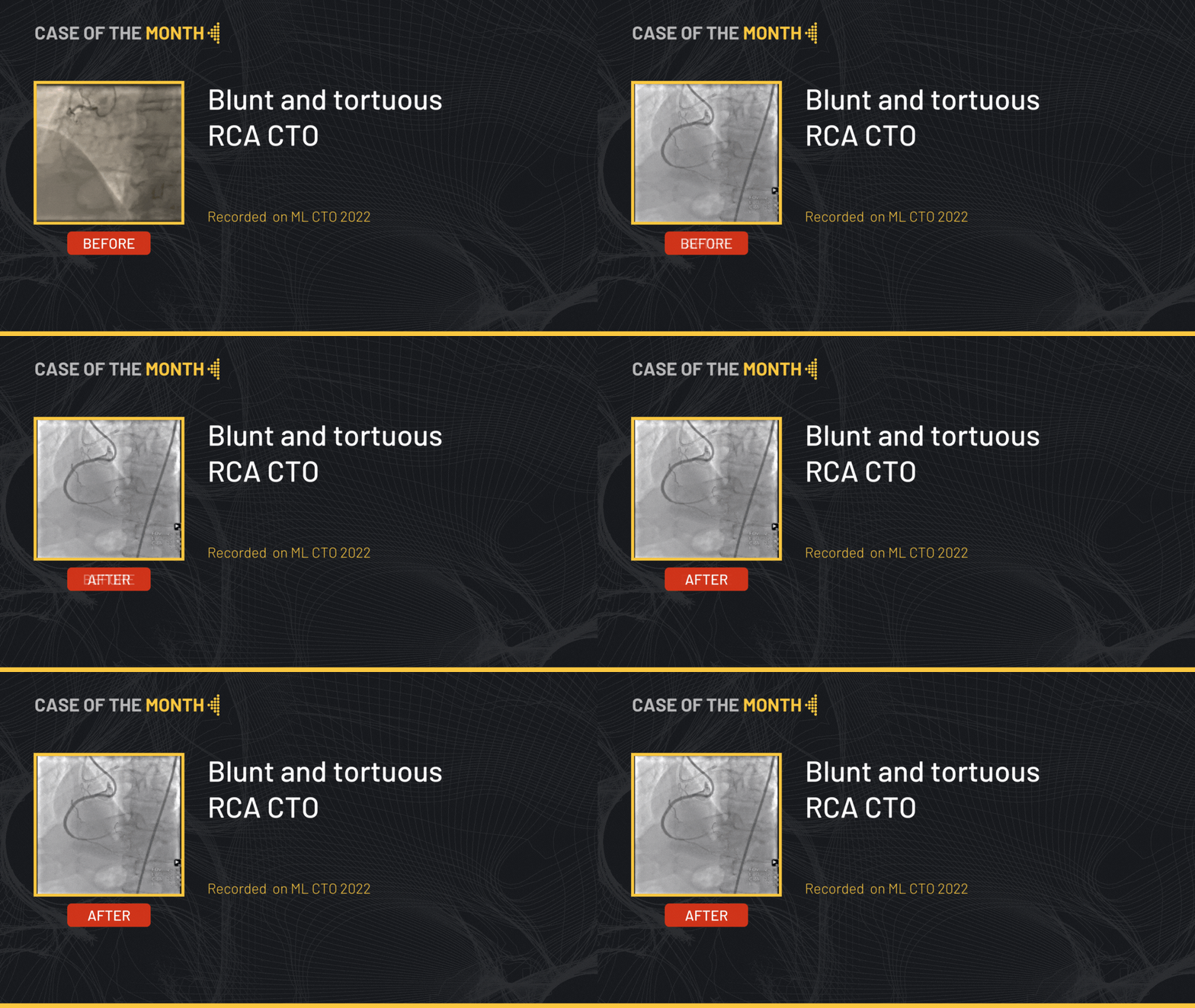
This month, we highlight the step by step approach of a tortuous RCA CTO lesion in a 71 years old male patient, s/p LAD stenting with ischemia at the level of the anterior and inferior walls on myocardial scintigraphy. The RCA shows a 20 mm calcified occlusion at the proximal level, with a relatively tapered proximal cap and retrograde filling from the septals.
Educational Objectives
- Plan a step-by-step approach procedure for CTO lesions.
- Antegrade wire escalation.
- Access sites and size possibilities during procedure.
- Wires to favor/avoid during subintimal space manipulation.
- IVUS role in CTO.
Step-by-step procedure:
1) Access site:
- Right radial approach: 7 French EBU to the left main + workhorse wire towards the distal LAD.
- Right femoral approach: 8 French AL to the RCA.
2) Step by step approach:
- The initial strategy was antegrade wire escalation.
- Using a Corsair Pro microcatheter a Fielder XT-R wire made progress through the CTO body advancing it further with the help of a retrograde injection.
- The wire appeared to be within the vessel architecture nonetheless following a deflecting trajectory.
- Escalation of the wire for a Gaia Second that finds a better position but was still subintimal.
- Redirecting the Gaia Second towards the intraluminal space followed by microcatheter advancement.
- De-escalation back to the Fielder XT-R that finds the intraluminal space.
- Exchange of the Fielder XT-R for a Sion blue wire followed by trapping and retrieving of microcatheter.
- Pre-dilatation of the occlusion body by a 3.0 mm balloon was performed.
- Stenting of the ostial RCA using 3.5x48 mm stent was followed by intravascular ultrasound (IVUS) which confirmed a long subintimal path after the distal edge of the stent with a typical image of an extraplaque path with a well apposed stent proximally.
- Additional stenting at the extraplaque level using a 3.5x20 mm stent was performed.
- The final angiographic end-result showed a satisfactory result.
Bibliography
1. Kalogeropoulos, A.S.; Alsanjari, O.; Davies, J.R.; Keeble, T.R.; Tang, K.H.; Konstantinou, K.; Vardas, P.; Werner, G.S.; Kelly, P.A.; Karamasis, G. V. Impact of Intravascular Ultrasound on Chronic Total Occlusion Percutaneous Revascularization. Cardiovasc. Revascularization Med. 2021, 33, 32–40, doi:10.1016/j.carrev.2021.01.008.
2. Xhepa, E.; Cassese, S.; Rroku, A.; Joner, M.; Pinieck, S.; Ndrepepa, G.; Kastrati, A.; Fusaro, M. Subintimal Versus Intraplaque Recanalization of Coronary Chronic Total Occlusions: Mid-Term Angiographic and OCT Findings From the ISAR-OCT-CTO Registry. JACC Cardiovasc. Interv. 2019, 12, 1889–1898, doi:10.1016/j.jcin.2019.04.049.
3. Denby, K.; Young, L.; Ellis, S.; Khatri, J. Antegrade Wire Escalation in Chronic Total Occlusions: State of the Art Review. Cardiovasc. Revasc. Med. 2023, 55, 88–95, doi:10.1016/J.CARREV.2023.06.011.
4. Maeremans, J.; Knaapen, P.; Stuijfzand, W.J.; Kayaert, P.; Pereira, B.; Barbato, E.; Dens, J. Antegrade Wire Escalation for Chronic Total Occlusions in Coronary Arteries: Simple Algorithms as a Key to Success. J. Cardiovasc. Med. 2016, 17, 680–686, doi:10.2459/JCM.0000000000000340
Date du tournage : 09/04/2024
Dernière mise à jour : 09/04/2024
Dernière mise à jour : 09/04/2024
Our Cases of the Month
The case of the month is a new way for our users to watch, learn, and share with incathlab. They can watch a video that highlights an innovative case and uses excellent pedagogical techniques, lear...
Partager
Suggestions
Protected Carotid Artery Stenting with a new micromesh carotid stent
Case of the month: November 2022
Partager
Calcified distal Left Main and LAD stenoses - Rotablation treatment and IVUS evalutation
Case of the month: June 2017
Partager


