×
Il semble que vous utilisiez une version obsolète de internet explorer. Internet explorer n'est plus supporté par Microsoft depuis fin 2015. Nous vous invitons à utiliser un navigateur plus récent tel que Firefox, Google Chrome ou Microsoft Edge.
My Player placeholder

Devenez membre d'Incathlab et bénéficiez d'un accès complet !

Vous devez être membre pour accéder aux vidéos Incathlab sans limitation. Inscrivez vous gratuitement en moins d'une minute et accédez à tous les services Incathlab ! Vous avez aussi la possibilité de vous connecter directement avec votre compte facebook ou twitter en cliquant sur login en haut à droite du site.

Inscription Connexion


This month, we highlight the case of ascending aortic pseudo aneurysm closure in a female patient, s/p CABG and aortic valve replacement as well as composite graft for an ascending aortic aneurysm, presenting with NSTEMI treated by stenting. A widened mediastinum was found on chest X-Ray leading to an angiography CT-scan showing a pseudo-aneurysm of the ascending aorta at the level of the previous sutures of the graft that was referred to percutaneous closure after surgical turn-down.

Educational Objectives

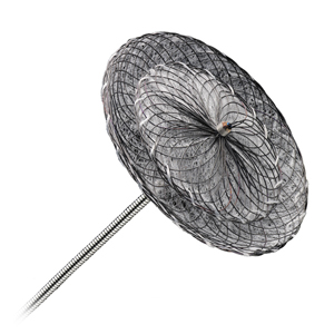
  • Learn how to prepare a VSD plug device.
  • Learn how to deploy a VSD plug device.
  • Learn the step-by-step approach to percutaneous closure of an aortic pseudo aneurysm.

Step-by-step procedure:

1) Access site:

  • 8 Fr bi-femoral approach.

2) Step by step approach:

An AL1 8Fr guiding catheter was not able to engage the pseudoaneurysm at first prompting the operators to perform an aortography using a pigtail catheter.

A 6Fr Judkins Right guiding catheter was also not able to engage despite trials using coronary guidewires (Whisper ES and Sion Black).

An AL1 6Fr guiding catheter found the false lumen and was exchanged for an 8Fr MP guiding cathter using a Confida wire.

The VSD plug (8Fr) was prepared and advanced over the stylet to the level of the aneurysm neck.

Pullback was performed after deployment of the distal end until resistance was felt after which an aortogram confirmed the correct position followed by full deployment of the device and retrieval of the guiding catheter.

A final aortogram was performed confirming the good sealing of the aneurysm.

Bibliography

1. ​Pirelli, L.; Kliger, C.; Fontana, G.P.; Ruiz, C.E.; Ruiz, C.E. Direct Percutaneous Transaortic Approach for Treatment of Aortic Pseudoaneurysms. 2015, doi:10.1093/icvts/ivv027.
 
2. Harky, A.; Khosravi, A.; Elam, L.; Colledge, J.; Kolvekar, S. Percutaneous Intervention of a Rare Complication Post Cardiac Surgery. 2017, doi:10.1016/j.athoracsur.2017.02.024.
 
3. ​​​Khanji, M.Y.; Kumar, P.; Ionescu, A. Ascending Aortic Pseudo-Aneurysm Treated with “Coil and Plug.” Eur. Hear. journal. Cardiovasc. Imaging 2017, 18, 1299, doi:10.1093/EHJCI/JEX179.
Date du tournage : 16/07/2018
Dernière mise à jour : 29/07/2024
Chris Zambakides
Germiston, Afrique Du Sud

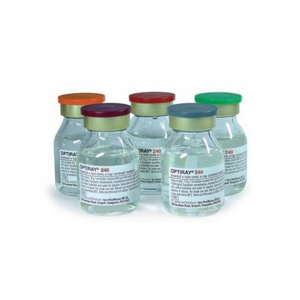
OptiRAY® / Guerbet

Optiray® contrast agent is lower osmolar, lower viscosity and nonionic.

Our Cases of the Month

The case of the month is a new way for our users to watch, learn, and share with incathlab. They can watch a video that highlights an innovative case and uses excellent pedagogical techniques, lear...

Partager
4 comments
Participer à la discussion
  • omer S. perfect.

      Sélectionnez vos fichiers, cliquez sur le bouton envoyer, rédigez votre comentaire puis cliquez sur le bouton envoyer. (formats autorisés : images jpeg, gif, png, and PDF)
      Your browser doesn't have Flash, Silverlight or HTML5 support.


    • Harun A. What do you think about putting some coils inside the sac before closing the neck. Because endoleak may persist while the patient using oral anticoagulant.

        Sélectionnez vos fichiers, cliquez sur le bouton envoyer, rédigez votre comentaire puis cliquez sur le bouton envoyer. (formats autorisés : images jpeg, gif, png, and PDF)
        Your browser doesn't have Flash, Silverlight or HTML5 support.


      • haldun T. why not putting in a 5 cm TEVAR extention since htere is enough proksimal graft length and adequate distance to the orifice of brachiocephalic artery??

          Sélectionnez vos fichiers, cliquez sur le bouton envoyer, rédigez votre comentaire puis cliquez sur le bouton envoyer. (formats autorisés : images jpeg, gif, png, and PDF)
          Your browser doesn't have Flash, Silverlight or HTML5 support.


        • Nayef Z. Super

            Sélectionnez vos fichiers, cliquez sur le bouton envoyer, rédigez votre comentaire puis cliquez sur le bouton envoyer. (formats autorisés : images jpeg, gif, png, and PDF)
            Your browser doesn't have Flash, Silverlight or HTML5 support.


          Suggestions

          Mardi 11 septembre 2018 de 12h30 à 14h (GMT+2)
          Honolulu : Mardi 11 septembre 2018 de 01h30 à 03h (GMT+2)
          San Francisco : Mardi 11 septembre 2018 de 04h30 à 06h (GMT+2)
          New York : Mardi 11 septembre 2018 de 07h30 à 09h (GMT+2)
          Buenos Aires : Mardi 11 septembre 2018 de 08h30 à 10h (GMT+2)
          Reykjavik : Mardi 11 septembre 2018 de 11h30 à 13h (GMT+2)
          London / Dublin : Mardi 11 septembre 2018 de 12h30 à 14h (GMT+2)
          Paris / Berlin : Mardi 11 septembre 2018 de 13h30 à 15h (GMT+2)
          Istanbul : Mardi 11 septembre 2018 de 14h30 à 16h (GMT+2)
          Moscou / Dubaï : Mardi 11 septembre 2018 de 15h30 à 17h (GMT+2)
          Bangkok : Mardi 11 septembre 2018 de 18h30 à 20h (GMT+2)
          Shanghai : Mardi 11 septembre 2018 de 19h30 à 21h (GMT+2)
          Tokyo : Mardi 11 septembre 2018 de 20h30 à 22h (GMT+2)
          Sydney : Mardi 11 septembre 2018 de 22h30 à 00h (GMT+2)
          Wellington : Mercredi 12 septembre 2018 de 00h30 à 02h (GMT+2)

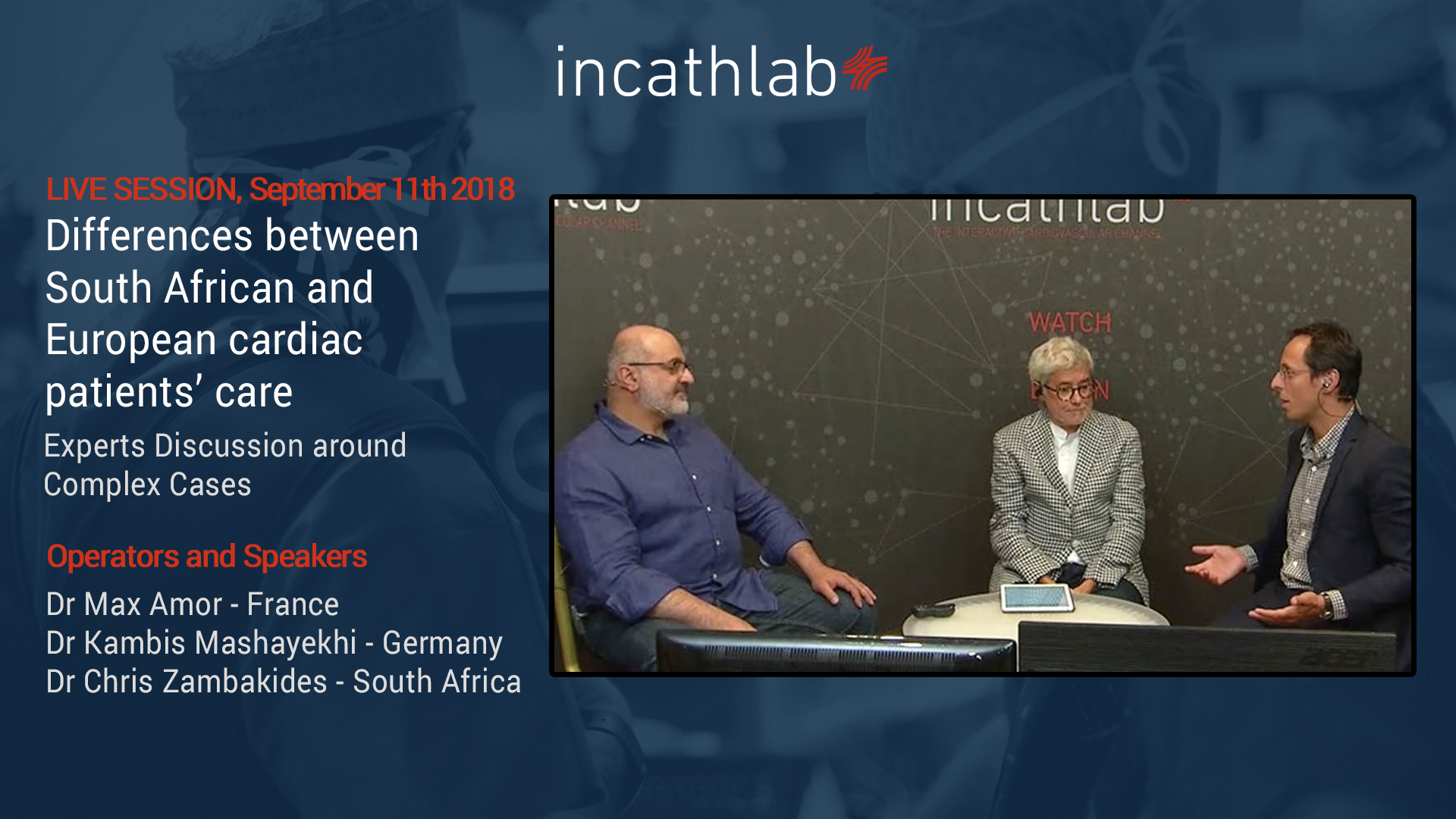
          Differences between South African and European cardiac patients’ care

          Experts Discussion around Complex Cases

          Partager
          Vendredi 29 juin 2018 de 09h40 à 11h (GMT+2)
          Honolulu : Jeudi 28 juin 2018 de 22h40 à 00h (GMT+2)
          San Francisco : Vendredi 29 juin 2018 de 01h40 à 03h (GMT+2)
          New York : Vendredi 29 juin 2018 de 04h40 à 06h (GMT+2)
          Buenos Aires : Vendredi 29 juin 2018 de 05h40 à 07h (GMT+2)
          Reykjavik : Vendredi 29 juin 2018 de 08h40 à 10h (GMT+2)
          London / Dublin : Vendredi 29 juin 2018 de 09h40 à 11h (GMT+2)
          Paris / Berlin : Vendredi 29 juin 2018 de 10h40 à 12h (GMT+2)
          Istanbul : Vendredi 29 juin 2018 de 11h40 à 13h (GMT+2)
          Moscou / Dubaï : Vendredi 29 juin 2018 de 12h40 à 14h (GMT+2)
          Bangkok : Vendredi 29 juin 2018 de 15h40 à 17h (GMT+2)
          Shanghai : Vendredi 29 juin 2018 de 16h40 à 18h (GMT+2)
          Tokyo : Vendredi 29 juin 2018 de 17h40 à 19h (GMT+2)
          Sydney : Vendredi 29 juin 2018 de 19h40 à 21h (GMT+2)
          Wellington : Vendredi 29 juin 2018 de 21h40 à 23h (GMT+2)

          Complex Right Coronary Artery CTO Procedure: Step-by-Step antegrade dissection reentry technique

          Case of the month: September 2018 - Live Case #6 MLCTO 2018

          Partager
          Scroll Up