×
It looks like you're using an obsolete version of internet explorer. Internet explorer is no longer supported by Microsoft since the end of 2015. We invite you to use a newer browser such as Firefox, Google Chrome or Microsoft Edge.
My Player placeholder

Become an Incathlab member and receive full access to its content!
You must be an Incathlab member to access videos without any restrictions. Register for free in one minute and access all services provided by Incathlab.You will also be able to log into Incathlab from your Facebook or twitter account by clicking on login on the top-right corner of Incathlab website.
Registration Login
Registration Login
This didactic procedure concerns a 72 years old women with a history of hypertension and Diabetes, she presented weight loss of 10 kilograms, distaste of meal and post prandial with pain one month before investigation.
CT scan showed: Complete occlusion of calcified celiac trunk with Severely calcified stenosed mesenteric artery. A collateral Riolan arc is present from inferior mesenteric artery to superior mesenteric artery. See CT figures
Educational objectives
- Plan a step-by-step procedure.
- How to select and brachial access.
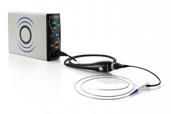
- Use of IVL before stenting
- How to proceed to a safe and successful positioning of the endoprosthesis?
- Materials choice.
- Tips and tricks for vessel preparation a good endoprosthesis positioning
- Endoprosthesis deployment.
Step-by-step procedure:
1) Vascular imaging: CT/angiographic imaging
- Identifying the localization and the extent of the lesion
- Identifying the adjacent vessels that could interfere with the stent
- Identifying access challenges: vessel stenosis, tortuosity, anatomy
2) Vascular access
- 1st access: right brachial access: Gentle navigation through the brachial artery and advance a 90cm 6 Fr braided sheath introducer
- Crossing of mesenteric stenosis with JR4 5F 125cm and a 300 cm 0.014” Guidewire
- Dilatation with IVL catheter
3) Mesenteric artery stenting
- Identifying the proximal and distal landing zone
- Crossing of mesenteric stenosis with JR4 5F 125cm and a 300 cm 0.014” Guidewire
- Wiring lesion with a 0.014 ” Spartacore
- Dilatation with IVL catheter
- Advancement of the balloon Athletis 5 x 4 x 135 cm with active control with gentle navigation
- Predilatation with 12 ATM
- Angiographic control after predilatation
- After predilatation : Positioning of the endoprosthesis.
- Important remarks to take into consideration while positioning the endoprosthesis:
- Angiographic control of the position of the endoprosthesis
4) Deployment of the endoprosthesis
- Opening of the endoprosthesis
- Angiographic control of the deployment and the position of the first struts
- Adjust the endoprosthesis position up/downstream for an optimal positioning
- Complete opening of the endoprosthesis from distal to proximal
- Complete deployment after release the angulation control
- Angiographic control of the final endoprosthesis position
5) Vascular closure with manual compression
6) Clinical observation and Follow-up in CCU for 24h.
7) CT scan after procedure showed : Excellent deployment of the stent . Minimal residual stenosis
Before

After


Bibliography
Shooting date : 2022-09-01
Last update : 2023-07-11
Last update : 2023-07-11
Our Cases of the Month
The case of the month is a new way for our users to watch, learn, and share with incathlab. They can watch a video that highlights an innovative case and uses excellent pedagogical techniques, lear...
Share
Join the Discussion
Suggestions
Monday, November 30th -0001 from 12am to 12am (GMT+1)
Honolulu : Monday, November 29th 1999 from 12pm to 12pm (GMT+1)
San Francisco : Monday, November 29th 1999 from 02pm to 02pm (GMT+1)
New York : Monday, November 29th 1999 from 05pm to 05pm (GMT+1)
Buenos Aires : Monday, November 29th 1999 from 07pm to 07pm (GMT+1)
London / Dublin : Monday, November 29th 1999 from 10pm to 10pm (GMT+1)
Paris / Berlin : Monday, November 29th 1999 from 11pm to 11pm (GMT+1)
Istanbul : Tuesday, November 30th 1999 from 12am to 12am (GMT+1)
Moscou / Dubaï : Tuesday, November 30th 1999 from 02am to 02am (GMT+1)
Bangkok : Tuesday, November 30th 1999 from 05am to 05am (GMT+1)
Shanghai : Tuesday, November 30th 1999 from 06am to 06am (GMT+1)
Tokyo : Tuesday, November 30th 1999 from 07am to 07am (GMT+1)
Sydney : Tuesday, November 30th 1999 from 08am to 08am (GMT+1)
Wellington : Tuesday, November 30th 1999 from 10am to 10am (GMT+1)
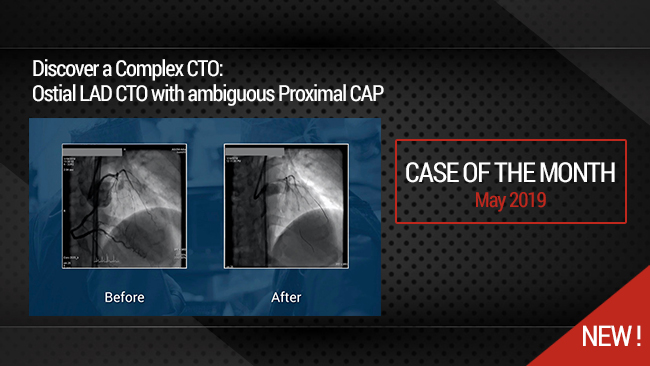
Complex CTO: Ostial LAD CTO with ambiguous Proximal CAP
Case of the month: May 2019
Share
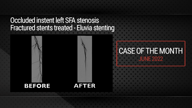
Occluded instent left SFA stenosis | Fractured stents treated - Eluvia stenting
Case of the month: June 2022
Share
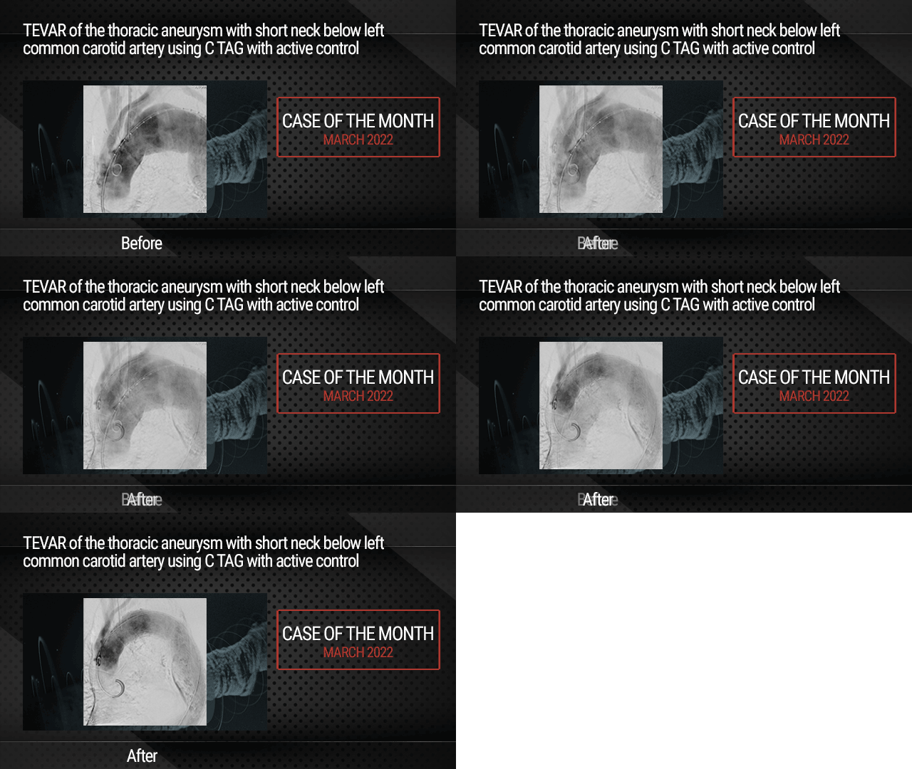
TEVAR of the thoracic aneurysm with short neck below left common carotid artery using C TAG with act...
Case of the month: March 2022
Share
Recanalization for limb salvage
Three occlusions: femoral, popliteal and posterior tibial arteries - Case of the month: December 201...
Share
Progressive Right Internal Carotid Stenosis with Left Internal Carotid Artery Occlusion
Case of the month: November 2017
Share


Ben jemaa H. :)