×
It looks like you're using an obsolete version of internet explorer. Internet explorer is no longer supported by Microsoft since the end of 2015. We invite you to use a newer browser such as Firefox, Google Chrome or Microsoft Edge.
My Player placeholder

Become an Incathlab member and receive full access to its content!
You must be an Incathlab member to access videos without any restrictions. Register for free in one minute and access all services provided by Incathlab.You will also be able to log into Incathlab from your Facebook or twitter account by clicking on login on the top-right corner of Incathlab website.
Registration Login
Registration Login
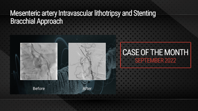
This didactic procedure concerns a 77 years old Women with diabetes , HBP, Dyslipidemia , presenting infra abdominal and lower limbs resting pain and bilateral trophic ulcers , due to distal abdominal aortic calcified occlusion . After multimodality assessment of lesions, she has undergone percutaneous abdominal aortic angioplasty and stenting with good final result.
Step-by-step procedure: How to deal with symptomatic Abdominal Aortic calcified occlusion
Educational objectives
- How to manage patients with lower limbs resting pain and abdominal Aortic occlusion
- Multimodality assessment of aortic occlusion before the intervention
- How to plan access, procedural steps, and selection of devices
- How to preserve renal artery and mesenteric artery during distal abdominal aortic occlusion
- How to prepare for stenting by Intra-Vascular Lithotripsy
1) Access sites:
- Brachial access : 6 French access using micro puncture system (COOK)
- Femoral access: 7 French access using micro puncture system (COOK)
- Heparin administration
2) Abdominal Aorta catheterization:
- Continuous flushing of the guiding catheter while introducing guidewire
- Advance softly an 6 Fr JR 4 guiding catheter to the descending aorta over a J 3 0.035’’ GUIDEWIRE
- Advance the 0.035” Guidewire towards the top of the aorta occlusion
- Advancing 70 cm 6F Braided SHEATH (Cook Flexor ® )
3) Crossing Aortic occlusion :
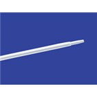
- With the support of the 6F catheter and a stiff angulated guidewire “0.018” Halberd wire Asahi®.
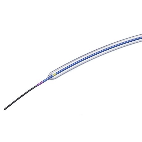
4) Pre-dilatation of the lesion with 6 x 80 mm Sterling balloon BSC
5) IVL Dilatation of the lesion with SHOCKWAVE balloon 8 mm x6cm , inflated to 4 ATM
6) Femoral retrograde recrossing of the aortic lesion using 0035’’ guidewire Advantage Terumo ® and BERENSTEIN 6F Catheter MERIT MEDICAL ®
7) Switching to Femoral introducer Sheath 12F , 45 cm Cook Flexor ®
8) Stenting
- Select the precise spot of stent deployment , in order to preserve collaterals . Control by brachial introducer
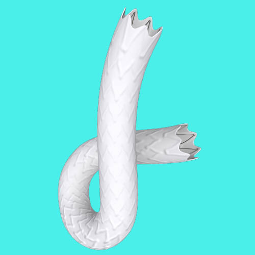
- Deployment of 14 x 49 mm Stent BeGraft Peripheral BENTLEY ® inflated to 5 ATM (two times)
- Control post stenting result , renal artery and mesenteric artery preserved
- Verify if there is any dissection or Rupture
9) Final angiographic control: ( 2 views Frontal & Lateral)
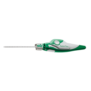
10) Vascular femoral closure with an 8 Fr Angio-Seal™
11) Medical adjunctive treatments
- Pre-procedural: Heparin , propofol and midazolam.
- Post procedural : double therapy: Aspirin 75mg o.d. + Clopidogrel 75mg o.d for one month
- After one month : Stop Clopidogrel and continue Aspirin 75mg
Bibliography
Shooting date : 2023-03-30
Last update : 2023-07-11
Last update : 2023-07-11
Our Cases of the Month
The case of the month is a new way for our users to watch, learn, and share with incathlab. They can watch a video that highlights an innovative case and uses excellent pedagogical techniques, lear...
Share
Join the Discussion
Suggestions
Complex Ostial LAD CTO: Saphenous vein Graft as an Option for Retrograde Approach
Case of the month: July 2019 - Live case #7 ML CTO
Share
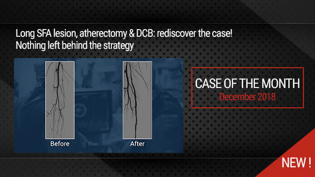
Nothing Left behind Strategy: Long SFA lesion, Directional atherectomy & DCB
Case of the month: December 2018
Share
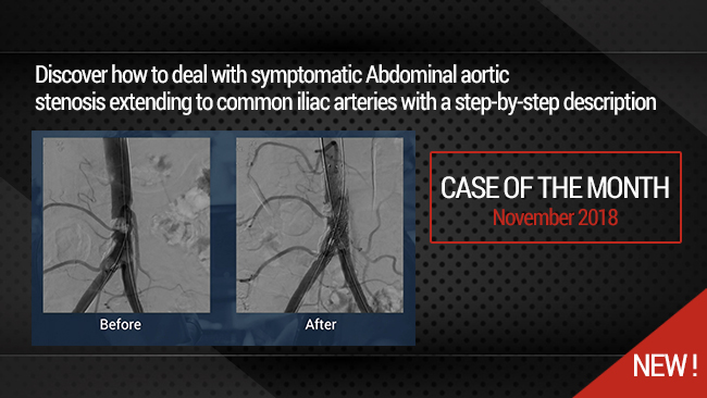
Complex Aorto-iliac lesion: How to deal with symptomatic Abdominal Aortic calcified stenosis extendi...
Case of the month: November 2018
Share
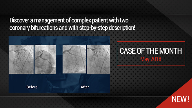
Simultaneous treatment of two coronary artery bifurcations in three vessels disease patient
Dedicated coronary bifurcation stents - Case of the month: May 2018
Share
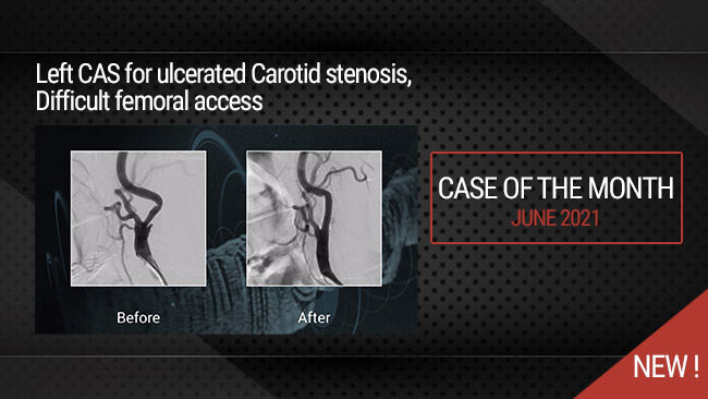
Left CAS for ulcerated Carotid stenosis, Difficult femoral access
Case of the month: June 2021
Share



Eli P. Thank you!