×
It looks like you're using an obsolete version of internet explorer. Internet explorer is no longer supported by Microsoft since the end of 2015. We invite you to use a newer browser such as Firefox, Google Chrome or Microsoft Edge.

Become an Incathlab member and receive full access to its content!
You must be an Incathlab member to access videos without any restrictions. Register for free in one minute and access all services provided by Incathlab.You will also be able to log into Incathlab from your Facebook or twitter account by clicking on login on the top-right corner of Incathlab website.
Registration Login
Registration Login
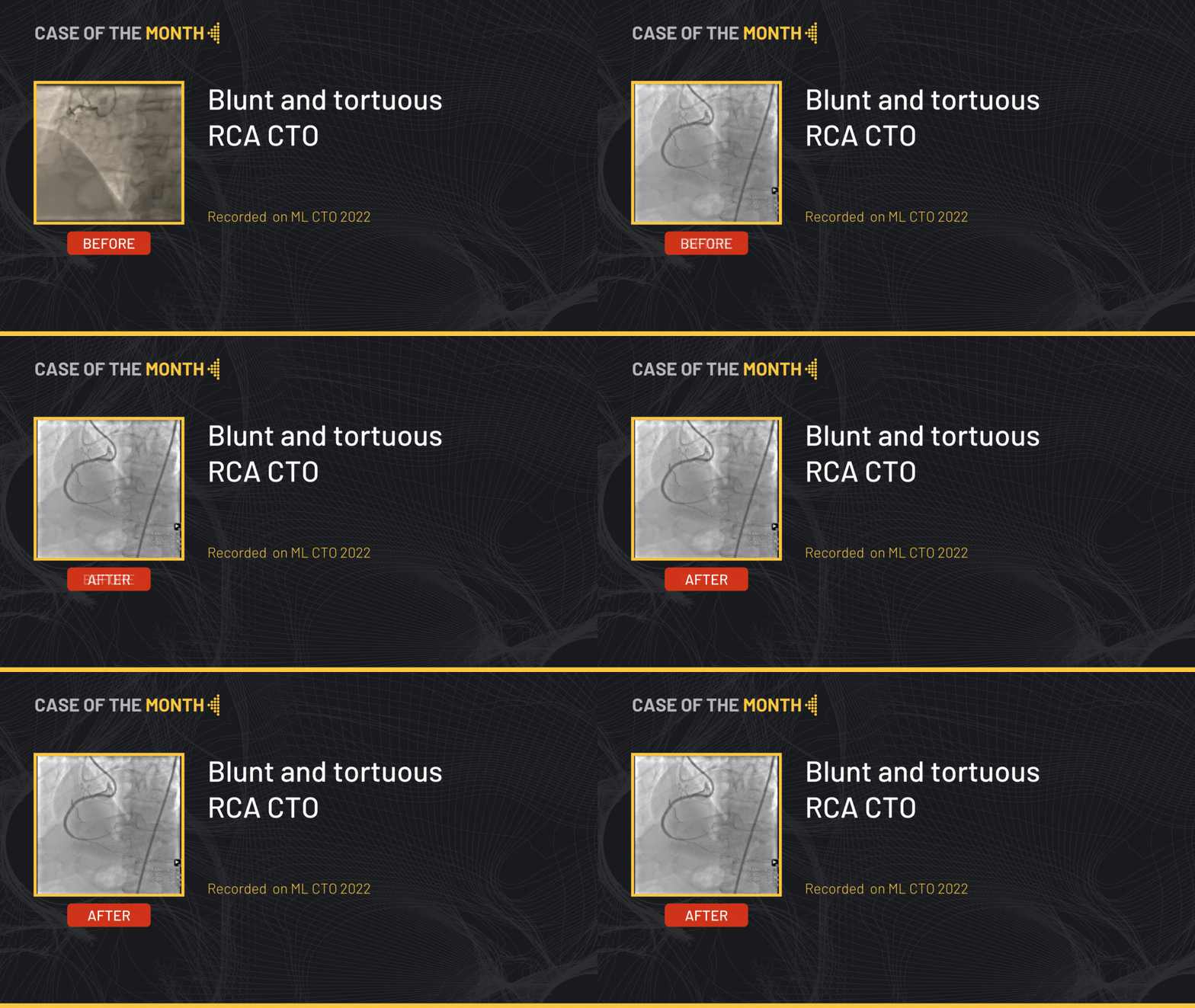
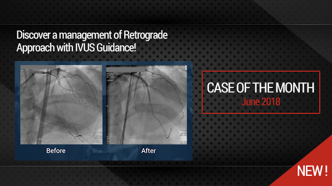
This month, we highlight the step by step approach of a tortuous RCA CTO lesion in a 71 years old male patient, s/p LAD stenting with ischemia at the level of the anterior and inferior walls on myocardial scintigraphy. The RCA shows a 20 mm calcified occlusion at the proximal level, with a relatively tapered proximal cap and retrograde filling from the septals.
Educational Objectives
- Plan a step-by-step approach procedure for CTO lesions.
- Antegrade wire escalation.
- Access sites and size possibilities during procedure.
- Wires to favor/avoid during subintimal space manipulation.
- IVUS role in CTO.
Step-by-step procedure:
1) Access site:
- Right radial approach: 7 French EBU to the left main + workhorse wire towards the distal LAD.
- Right femoral approach: 8 French AL to the RCA.
2) Step by step approach:
- The initial strategy was antegrade wire escalation.
- Using a Corsair Pro microcatheter a Fielder XT-R wire made progress through the CTO body advancing it further with the help of a retrograde injection.
- The wire appeared to be within the vessel architecture nonetheless following a deflecting trajectory.
- Escalation of the wire for a Gaia Second that finds a better position but was still subintimal.
- Redirecting the Gaia Second towards the intraluminal space followed by microcatheter advancement.
- De-escalation back to the Fielder XT-R that finds the intraluminal space.
- Exchange of the Fielder XT-R for a Sion blue wire followed by trapping and retrieving of microcatheter.
- Pre-dilatation of the occlusion body by a 3.0 mm balloon was performed.
- Stenting of the ostial RCA using 3.5x48 mm stent was followed by intravascular ultrasound (IVUS) which confirmed a long subintimal path after the distal edge of the stent with a typical image of an extraplaque path with a well apposed stent proximally.
- Additional stenting at the extraplaque level using a 3.5x20 mm stent was performed.
- The final angiographic end-result showed a satisfactory result.
Bibliography
1. Kalogeropoulos, A.S.; Alsanjari, O.; Davies, J.R.; Keeble, T.R.; Tang, K.H.; Konstantinou, K.; Vardas, P.; Werner, G.S.; Kelly, P.A.; Karamasis, G. V. Impact of Intravascular Ultrasound on Chronic Total Occlusion Percutaneous Revascularization. Cardiovasc. Revascularization Med. 2021, 33, 32–40, doi:10.1016/j.carrev.2021.01.008.
2. Xhepa, E.; Cassese, S.; Rroku, A.; Joner, M.; Pinieck, S.; Ndrepepa, G.; Kastrati, A.; Fusaro, M. Subintimal Versus Intraplaque Recanalization of Coronary Chronic Total Occlusions: Mid-Term Angiographic and OCT Findings From the ISAR-OCT-CTO Registry. JACC Cardiovasc. Interv. 2019, 12, 1889–1898, doi:10.1016/j.jcin.2019.04.049.
3. Denby, K.; Young, L.; Ellis, S.; Khatri, J. Antegrade Wire Escalation in Chronic Total Occlusions: State of the Art Review. Cardiovasc. Revasc. Med. 2023, 55, 88–95, doi:10.1016/J.CARREV.2023.06.011.
4. Maeremans, J.; Knaapen, P.; Stuijfzand, W.J.; Kayaert, P.; Pereira, B.; Barbato, E.; Dens, J. Antegrade Wire Escalation for Chronic Total Occlusions in Coronary Arteries: Simple Algorithms as a Key to Success. J. Cardiovasc. Med. 2016, 17, 680–686, doi:10.2459/JCM.0000000000000340
Shooting date : 2024-04-09
Last update : 2024-04-09
Last update : 2024-04-09
Our Cases of the Month
The case of the month is a new way for our users to watch, learn, and share with incathlab. They can watch a video that highlights an innovative case and uses excellent pedagogical techniques, lear...
Share
Suggestions
Monday, November 30th -0001 from 12am to 12am (GMT+1)
Honolulu : Monday, November 29th 1999 from 01pm to 01pm (GMT+1)
San Francisco : Monday, November 29th 1999 from 03pm to 03pm (GMT+1)
New York : Monday, November 29th 1999 from 06pm to 06pm (GMT+1)
Buenos Aires : Monday, November 29th 1999 from 08pm to 08pm (GMT+1)
London / Dublin : Monday, November 29th 1999 from 11pm to 11pm (GMT+1)
Paris / Berlin : Tuesday, November 30th 1999 from 12am to 12am (GMT+1)
Istanbul : Tuesday, November 30th 1999 from 01am to 01am (GMT+1)
Moscou / Dubaï : Tuesday, November 30th 1999 from 03am to 03am (GMT+1)
Bangkok : Tuesday, November 30th 1999 from 06am to 06am (GMT+1)
Shanghai : Tuesday, November 30th 1999 from 07am to 07am (GMT+1)
Tokyo : Tuesday, November 30th 1999 from 08am to 08am (GMT+1)
Sydney : Tuesday, November 30th 1999 from 09am to 09am (GMT+1)
Wellington : Tuesday, November 30th 1999 from 11am to 11am (GMT+1)
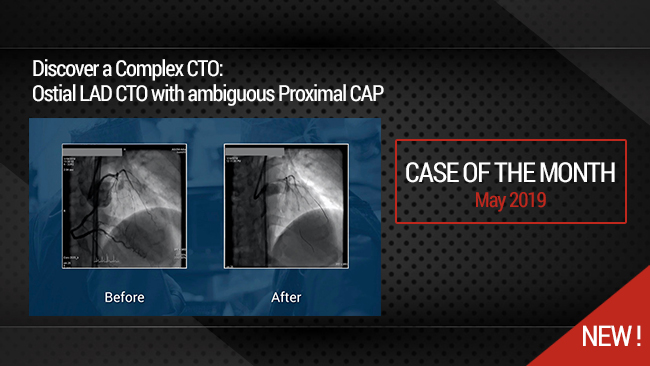
Complex CTO: Ostial LAD CTO with ambiguous Proximal CAP
Case of the month: May 2019
Share
Progressive Right Internal Carotid Stenosis with Left Internal Carotid Artery Occlusion
Case of the month: November 2017
Share


