×
It looks like you're using an obsolete version of internet explorer. Internet explorer is no longer supported by Microsoft since the end of 2015. We invite you to use a newer browser such as Firefox, Google Chrome or Microsoft Edge.
Complex Acute Anterior STEMI with "no Reflow phenomenon" management
Case of the Month: October 2018
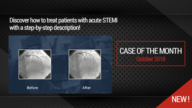
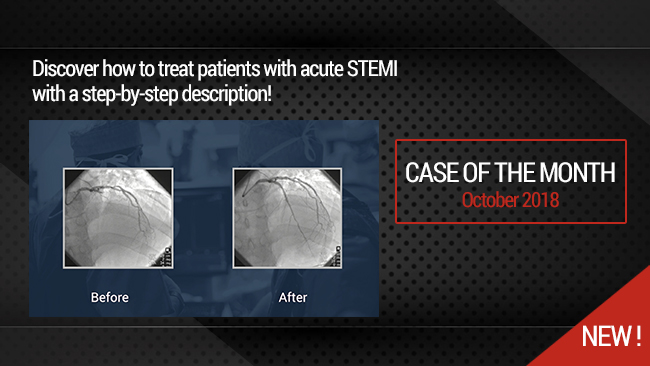
This didactic procedure concerns a patient adressed from neighboring hospital for acute anterior STEMI within the first hour. The coronary angiography has shown distal left main stenosis and Proximal LAD thrombotic occlusion.
This procedure show how to deal wih crossing difficulties during primary PCI as well as management of "no reflow phenomenon".
Educational objectives
- How to treat patients with acute STEMI.
- Some acute thrombotic lesions may be challenging to cross.
- How to use dedicated CTO devices during non-CTO PCI.
- How to use thrombo-aspiration catheter to deliver adenosine distally.
- How to treat "no reflow phenomenon" during primary PCI.
- IVUS guidance to control stent deployment & Left main stenting during primary PCI.
Step-by-Step procedure
- The patient has experienced repetitive cardiac arrest due to ventricular fibrillation, so he was intubated & admitted to cath-lab.
- Right radial 6F access.
- First coronary angiography: right system first.
- EBU 3.5 6F guiding catheter used for the left system.
- Left system angiography showed distal Left main significant lesion & proximal LAD thrombotic occlusion.
- The Sion black guidewire alone has failed to cross the lesion.
- Conventional balloon support (Rapid Exchange) has also failed to facilitate crossing the lesion.
- Finally the lesion was crossed succesfully with Sion black guidewire & CTO dedicated microcatheter (Turnpike LP: Teleflex) support.
- Predilatation with 2.0x20mm balloon has been performed.
- First stent implantation: Xience Sierra 3.0x28mm (Abbott) in the proximal-Mid LAD inflated at 12ATM.
- The angiographic control revealed LAD "non reflow ".
- Thrombo-aspiration cathter Export 6F (Medtronic) was used to deliver distally repetitive Adenosine Bolus.
- The angiographic control with Tip injection through the Export catheter showed sgnificant flow improvement.
- Second short stent Xience Sierra 2.75x8mm(Abbott) was implanted to cover distal edge dissection.
- IVUS was used to control LAD stents deployment & assessment of the distal Left main stenosis.
- Third Xience Sierra (abbott) 4.0x28mm was implanted on the left main to the Proximal LAD with some plaque shift to the Left circumflexe artery.
- POT was performed with a 5.0x8mm balloon, the patient has experienced again a Ventricular fibrillation during balloon infation.
- The final angiographic & hemodynamic results were satisfactory.
Protocol
- Contraste medium: Optiray 350 (Guerbet): 179ml.
- Prcedural time: 60min.
- Exposure time: 19min.
- Exposure: 2399mGy.
Biobliography
-
Acute Myocardial Infarction in patients presenting with ST-segment elevation (Management of) ESC 2017 Clinical Practice Guidelines - Article
Authors: Borja Ibanez (Chairperson) (Spain)
Publication: doi:10.1093/eurheartj/ehx393
-
Advances in Coronary No-Reflow Phenomenon-a Contemporary Review- Article
Authors:Karimianpour A, Maran A.
Publication: 2018 Jul 5;20(9):44. doi: 10.1007/s11883-018-0747-5.
-
Prediction of no-reflow and major adverse cardiovascular events with a new scoring system in STEMI patients.Link title - Article
Authors:Bayramoglu A, Ta§olar H, Kaya A, Tanboga 1H, Yaman M, Bekta§ 0, GUnaydin ZY, Oduncu V.
Publication: 2018 Apr;31(2):144-149. doi: 10.1111/joic.12463.
-
Role of Intravascular Ultrasound in Patients with Acute Myocardial Infarction - Article
Authors: Young Joon Hong, MD, Youngkeun Ahn, MD, and Myung Ho Jeong, MD
Publication: Korean Circ J. 2015 Jul; 45(4): 259-265.


